

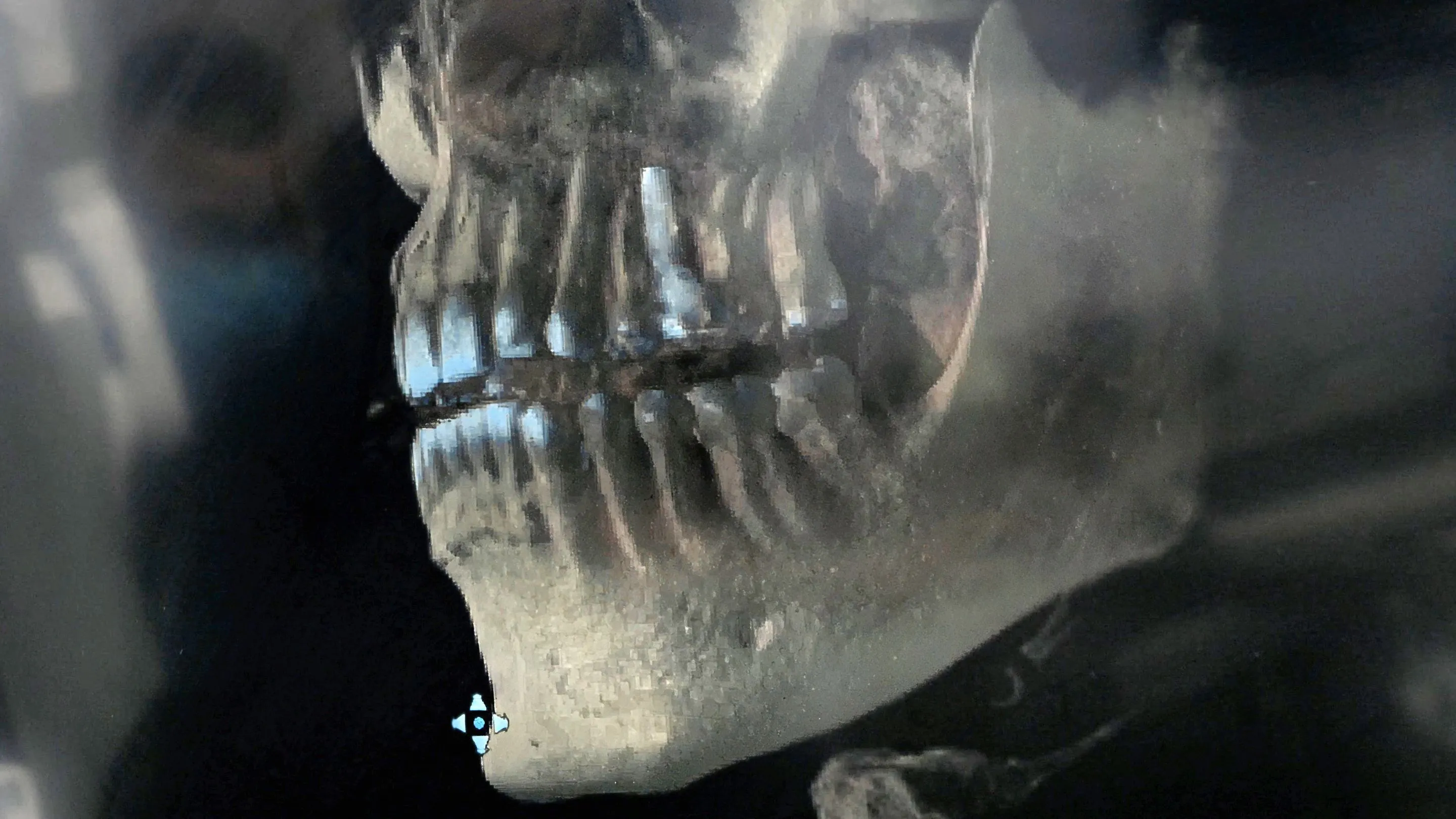
What cone beam imaging is
Cone beam computed tomography — usually shortened to CBCT or cone beam — rotates around the head once and captures a three-dimensional volume rather than a flat picture. The result can be turned, sliced and viewed from any angle on a screen.
A conventional X-ray flattens everything into a single plane, so structures that sit in front of and behind one another overlap. In 3D the dentist can see exactly where a nerve canal runs, how much bone is actually present and how close a root sits to the sinus — measurements rather than estimates.
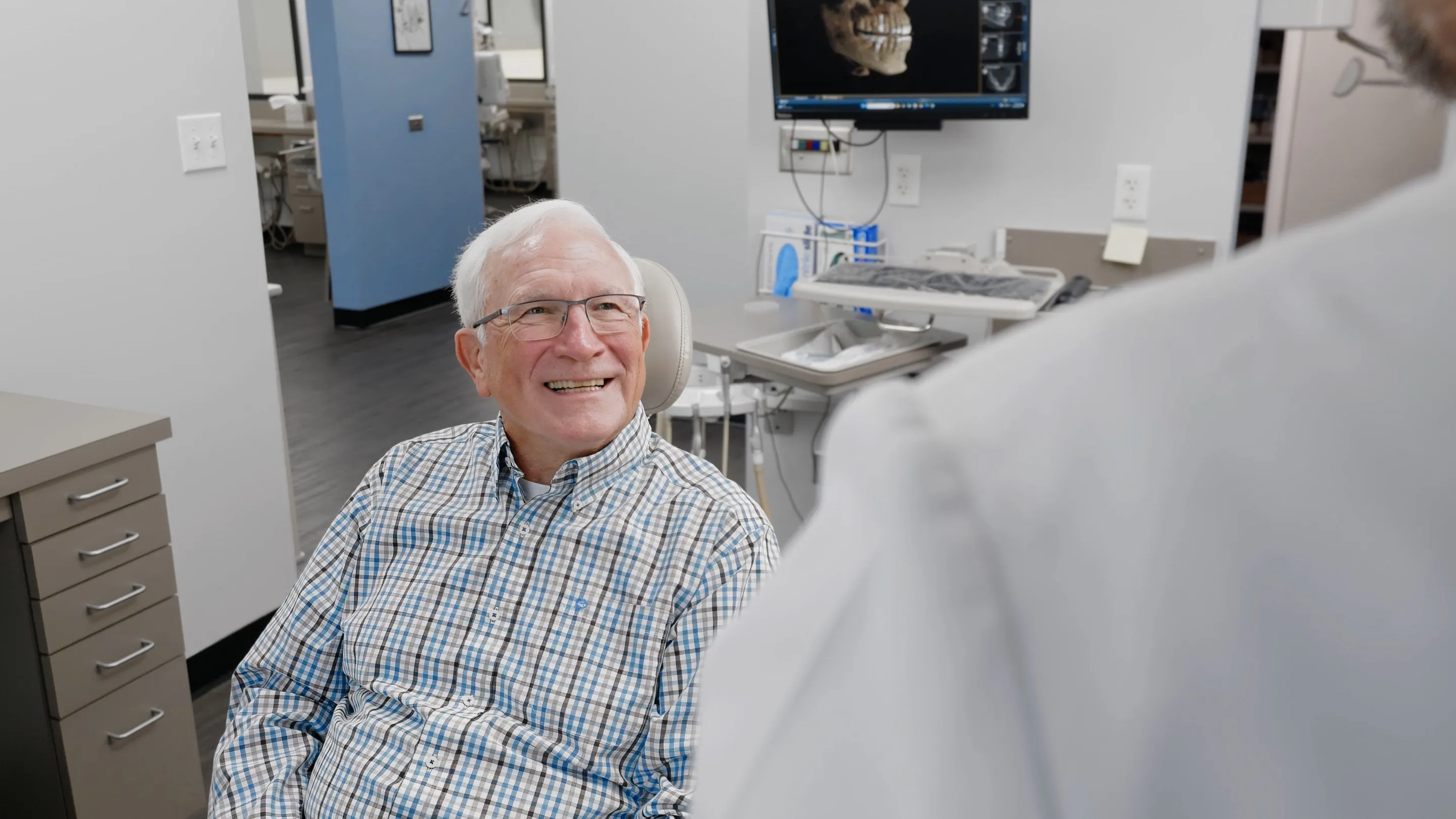
Compared with a two-dimensional X-ray, a cone beam scan can show:
- The height, width and density of the jawbone in a specific spot.
- The path of the nerve that runs through the lower jaw, and the floor of the sinus above the upper back teeth.
- The exact position of an impacted tooth and how close it sits to neighboring roots and nerves.
- Root shapes and extra canals that are hard to see on a flat image.
- Infection, cysts or other changes in the bone, and their true size.
- The bony parts of the jaw joint.
Most of that matters because it removes guesswork before a procedure, rather than discovering something part-way through one.
When it is used
Most often for planning dental implants, where the height, width and density of available bone decide whether an implant is possible, what size it should be and where it can safely go.
It is also used to assess impacted wisdom teeth and other teeth that have not come through, to plan extractions close to a nerve or the sinus, to investigate complicated root anatomy or a suspected crack ahead of root canal treatment, and to look at the jaw joint.
For a routine check-up it is not needed. Digital X-rays remain the right tool for finding decay and monitoring bone levels, with a lower dose.

The scan itself
You will be asked to remove anything metal around the head and neck — earrings, glasses, hairpins, removable dentures or partials — because metal causes streaks on the image.
You then sit or stand in the machine, usually resting your chin on a support and sometimes biting gently on a guide to keep the jaw in position. The arm rotates once around your head. The scan itself takes well under a minute and nothing touches you; the only thing asked of you is to stay still and breathe normally.
The images are ready to view on a screen shortly afterward, where the dentist can walk you through what they show.
A cone beam scan uses more radiation than a single conventional dental X-ray but generally considerably less than a conventional medical CT scan of the head. The dose also depends on how large an area is scanned: a scan limited to one part of the jaw uses less than one of the whole face.
That is why it is used selectively. The principle across dentistry is to keep radiation as low as reasonably achievable — scan only when a 3D view will change the plan, and scan only the area that needs it. If you are or might be pregnant, say so before any imaging.
Frequently Asked Questions
Is it uncomfortable?
No. Nothing is placed in your mouth apart from, sometimes, a small bite guide, and the machine does not touch you as it rotates. The main requirement is holding still for the few seconds the scan takes.
How long does it take?
The scan itself takes well under a minute — often less than half of that. Positioning you first takes a little longer than the scan.
Why not use it for everything?
Because the radiation dose is higher than a routine dental X-ray. Guidance across dentistry is to use cone beam imaging where a 3D view will genuinely change the diagnosis or the treatment plan, and to use ordinary X-rays where they are enough.
Is it the same as a CT scan at a hospital?
It works on a similar principle but is a different machine, designed for the teeth and jaws. It captures its image in one rotation and generally uses considerably less radiation than a conventional medical CT of the head.
Do I need one for a dental implant?
A 3D scan is widely used in implant planning because it shows the height, width and density of the bone and the position of the nerve and sinus, which a flat X-ray cannot. Whether one is needed in your case is decided at the consultation.
Can I have a cone beam scan if I am pregnant?
Tell the dental team if you are or might be pregnant. Imaging that is not urgent is often postponed, and where it is needed the decision is made with that in mind.