

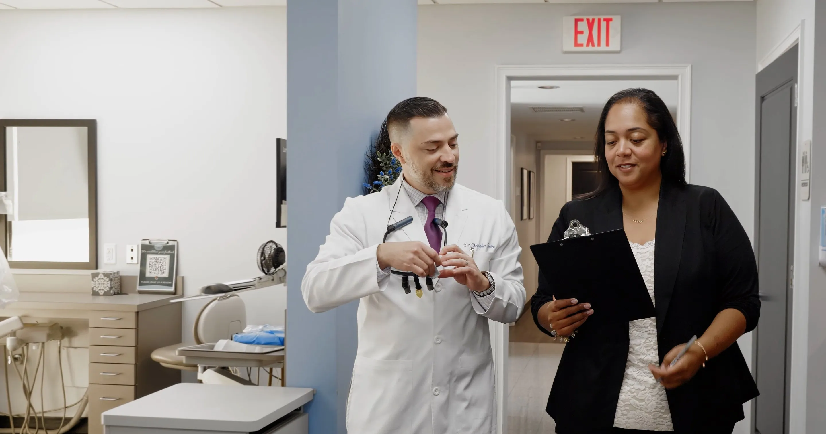
Why the equipment matters
Most dental technology exists to do one of three things: see more clearly, expose you to less radiation, or replace something uncomfortable with something that is not.
Seeing more clearly is the one that matters most. Much of what goes wrong in a mouth starts where it cannot be seen with the eye alone — between teeth where they touch, under an old filling, at the tip of a root, or in the bone. Better images mean problems are found earlier and treatment is planned more precisely, and found earlier usually means a smaller, simpler and less expensive fix.
The second benefit is less obvious but just as real: images on a screen can be shown to you. A crack, a shadow on an X-ray or the amount of bone available for an implant is much easier to make a decision about when you can see it for yourself.

What we use
Four kinds of imaging and scanning, each with its own job. Each page explains what it is, when it is used and what it is like.
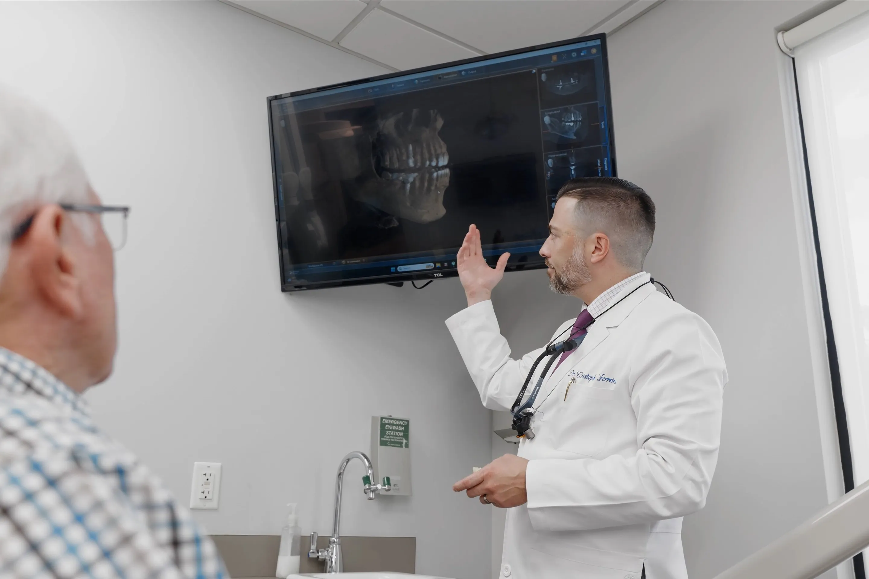
Digital X-Rays
Images on screen in seconds, with substantially less radiation than film — for decay between teeth, bone levels and infection at the root.
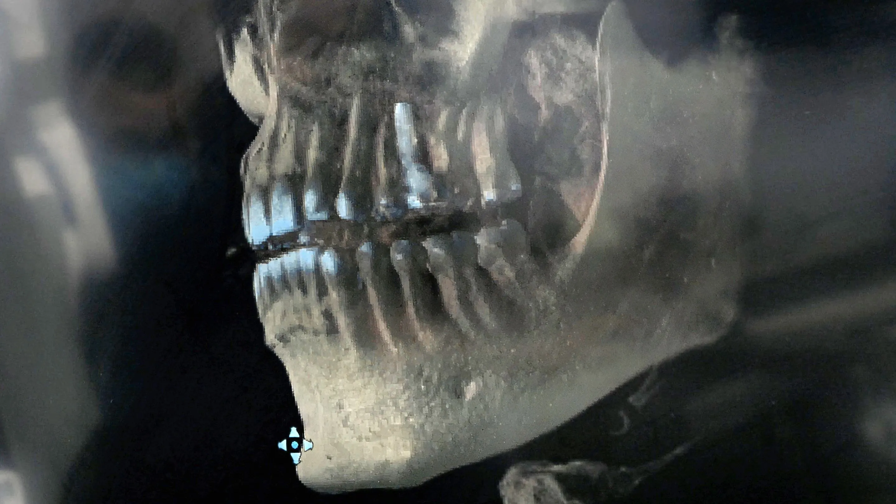
Cone Beam Imaging
A 3D scan of the jaw and teeth that shows bone, nerves and sinuses from any angle — used where a flat X-ray is not enough, such as implant planning.
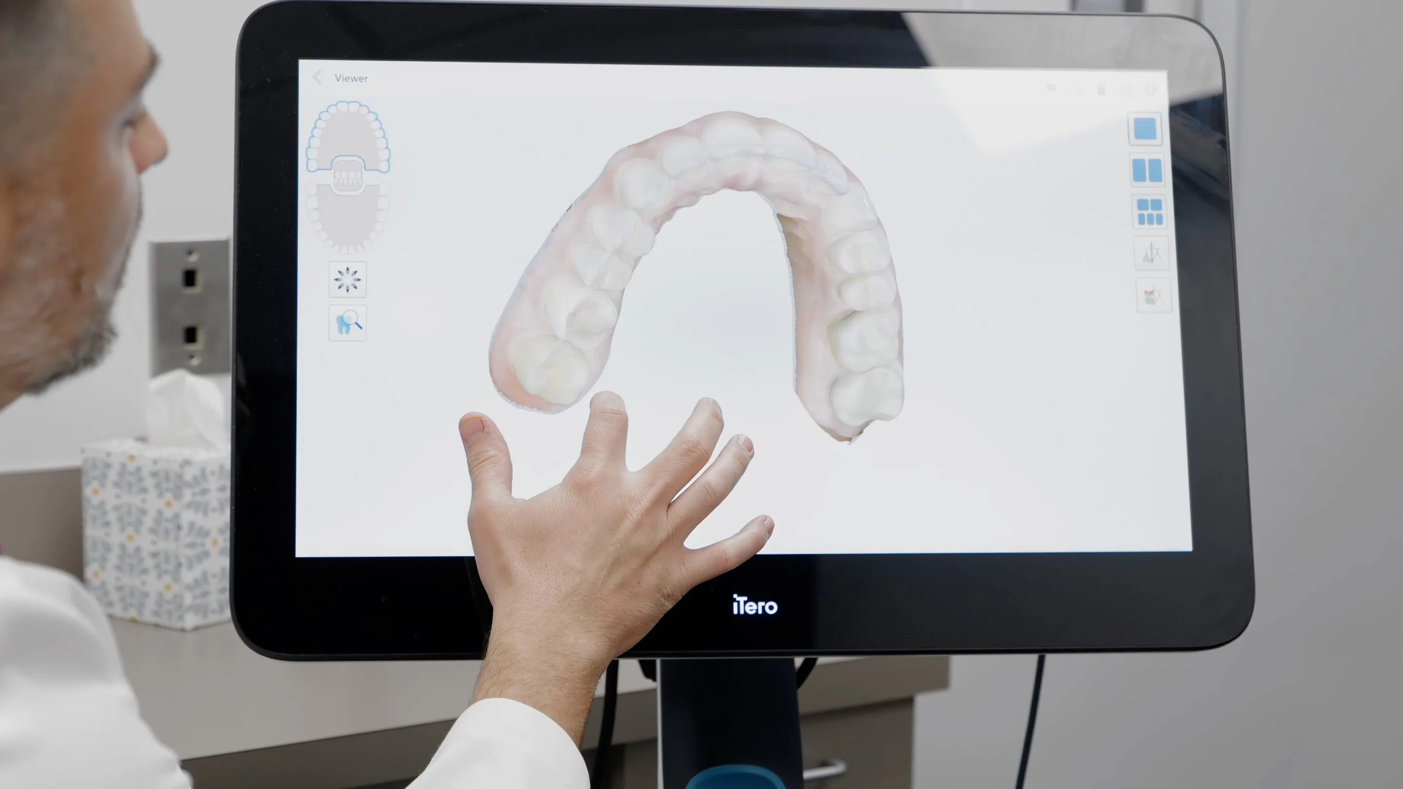
Digital Scanner
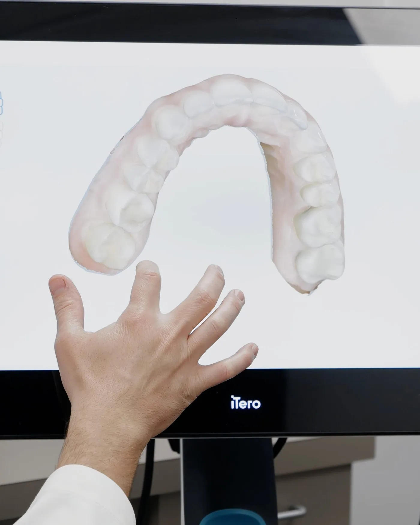
A small wand that builds a 3D model of your teeth on screen, in place of a tray of impression putty.

Intraoral Camera
A pen-sized camera that puts a magnified photograph of your own tooth on a screen, so you see what the dentist sees.
How the pieces fit together
The tools are rarely used on their own. A routine check-up might combine digital X-rays to look between the teeth and at the bone with an intraoral camera to show you a worn filling up close.
Planning an implant typically starts with a cone beam scan to measure the bone and locate the nerve and sinus, while a digital scan records the teeth and gums the final crown has to fit against. A crown, a night guard or a set of clear aligners can all begin with a digital scan instead of a tray of impression putty.
None of it replaces the examination itself. Images show what is there; it still takes a dentist to decide what it means and whether it needs doing anything about.
The part you do not see
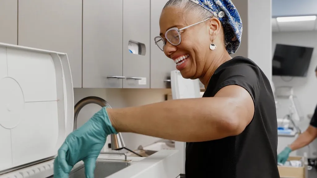
Not all of the equipment in a dental office is in the treatment room. In dentistry generally, reusable instruments are cleaned and sterilized between patients and single-use items are used once and discarded — infection-control guidance from the Centers for Disease Control and Prevention sets out how.
It is the least visible part of a visit and one of the most important. If you have questions about how instruments are handled, it is a perfectly reasonable thing to ask.
Frequently Asked Questions
Are dental X-rays safe?
The dose from routine dental X-rays is very small — a fraction of the natural background radiation everyone receives from the environment over a year — and digital sensors need less of it than film did. X-rays are taken when the information is needed for diagnosis, not on a fixed schedule for everyone.
Is a cone beam scan the same as a hospital CT scan?
It is related but not the same. A cone beam scanner is built for the teeth and jaws, captures its image in a single rotation, and generally uses considerably less radiation than a conventional medical CT of the head. It still uses more than a routine dental X-ray, so it is used where a 3D view changes the plan.
Will I have X-rays and a scan at every visit?
No. How often X-rays are taken depends on your own history and risk of decay and gum disease. A cone beam scan is only taken for specific planning or diagnostic questions, and a digital scan only when something is being made to fit your teeth.
Does a digital scanner replace every impression?
For many treatments it can, including most crowns, night guards and clear aligners. Some cases are still better suited to a conventional impression, and the dentist will choose whichever gives the more accurate result for the work being done.
Can I see my own images?
X-rays, scans and intraoral photographs all appear on a screen beside the chair, and being shown them is a normal part of the conversation about what was found. Ask if you would like something explained again.