


Seeing what the dentist sees
An intraoral camera is a pen-sized camera with its own light that takes magnified, sharp photographs inside the mouth and displays them on a screen beside the chair.
It changes the conversation. A cracked filling or a fractured cusp described in words is abstract, and a hand mirror shows very little of the back teeth. The same thing magnified many times on a screen is obvious, and it is much easier to decide about treatment when you can see the reason for it yourself.
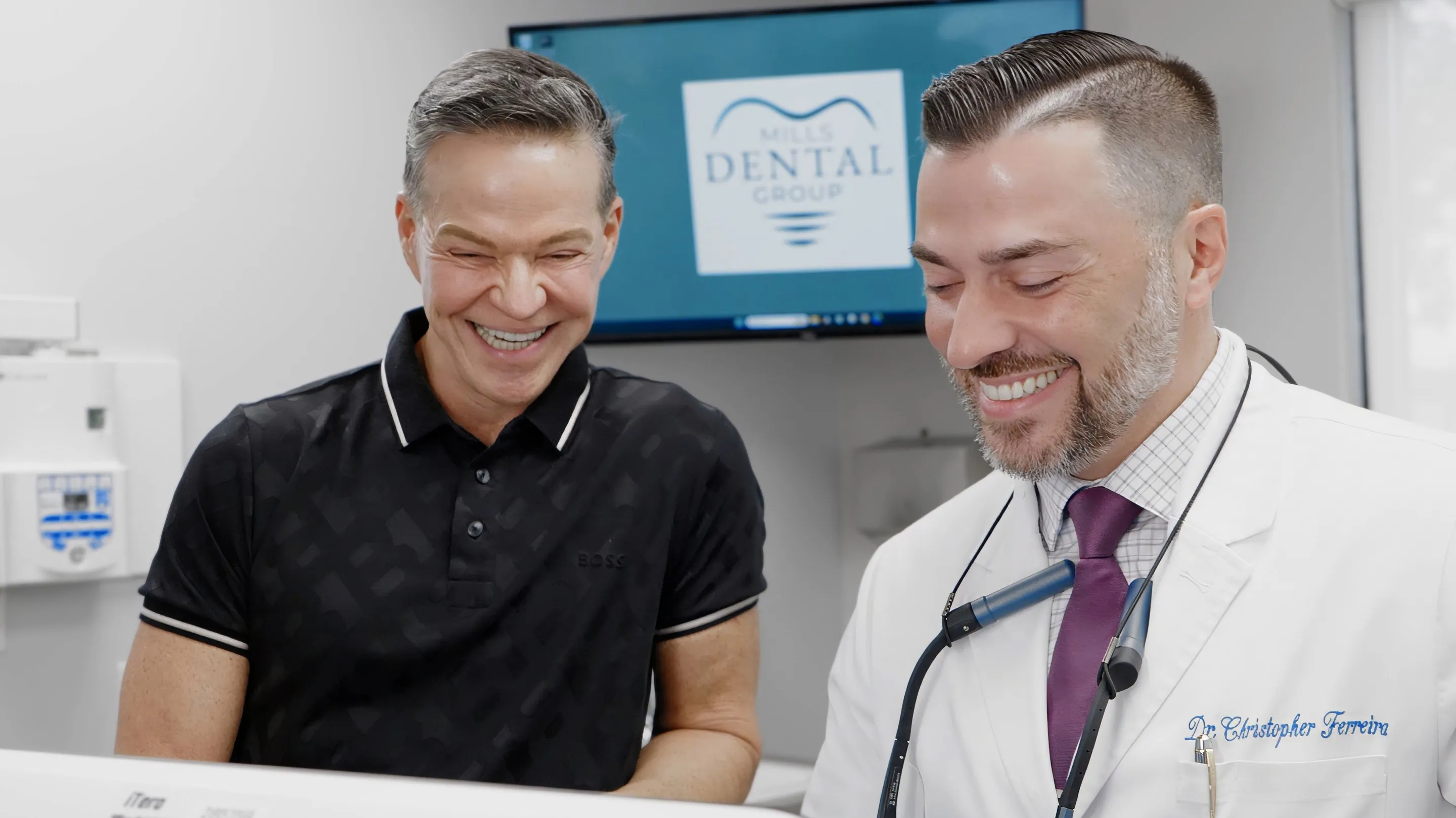
The camera is good at the things that are on the surface but hard to see:
- Cracks in teeth and fractures in old fillings.
- Worn or chipped edges, including wear from grinding.
- Decay that is visible on a biting surface or at the gumline.
- Gaps and leaks at the edges of fillings and crowns.
- Inflamed or receding gums, and areas where plaque or tartar builds up.
- Changes in the soft tissue of the cheeks, tongue and palate worth keeping an eye on.
What it cannot do is see inside a tooth, between teeth where they touch, or into the bone. That is what digital X-rays are for, and the two are often used together.
How it is used during a visit
The camera is usually picked up during an examination, when something is worth a closer look. The tip is moved to the tooth, the image appears on the screen straight away, and it can be frozen and enlarged while it is explained to you.
Nothing is placed in your mouth for more than a moment, and there is no preparation. If a problem is found — a crack that may need a crown, or a failing edge on an old filling — you will have seen it, rather than being asked to take it on trust.

Other uses
The images become part of your record, so a questionable area can be photographed and compared at the next visit to see whether it has changed. For something being watched rather than treated, that comparison is often the deciding factor.
They are also useful for insurance documentation, where a photograph can show why a treatment is needed, and for showing areas that are difficult to keep clean so that brushing and flossing can be adjusted. Before-and-after images give a clear record of what was done.
Frequently Asked Questions
Does it hurt?
No. It is a camera — it only takes pictures. The tip is small and smooth, and it rests against or just above the tooth for a moment while the image is captured.
Does an intraoral camera use radiation?
No. It uses ordinary light, like any other camera. It shows the surfaces of teeth and gums; X-rays are still needed to see between teeth and below the gum.
Is the camera clean?
Intraoral cameras are normally used with a disposable sleeve over the part that goes in the mouth, changed for every patient, and the handpiece is disinfected between uses.
Why photograph something that does not need treatment yet?
So it can be compared next time. A small crack or a worn edge that is being watched is much easier to judge against a clear photograph from six months ago than against a note in a chart.
Will I be asked to decide on treatment straight away?
No. The images are there to help you understand what was found. You can take time to think, ask questions and decide afterward.