

What changes with digital
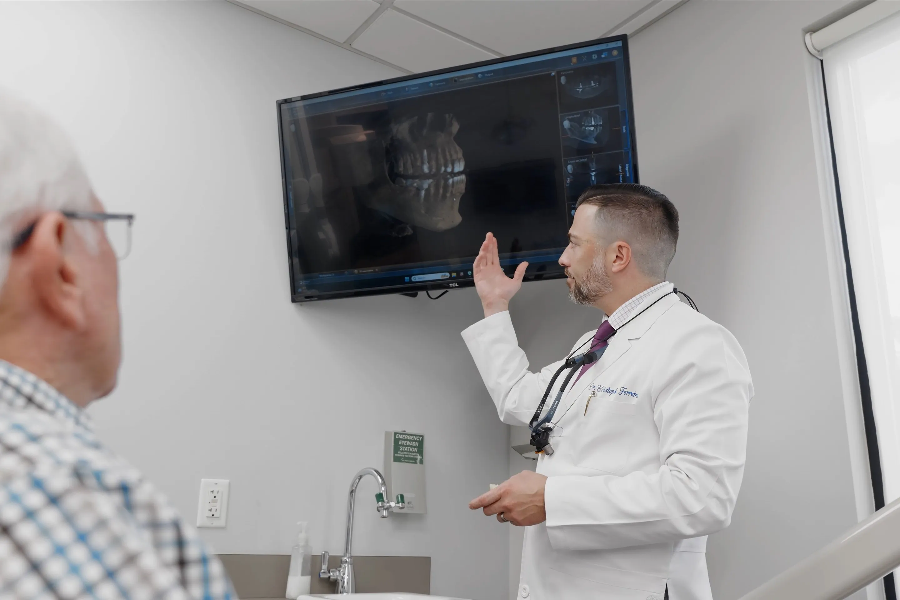
A digital sensor replaces the film packet. The image appears on a screen in seconds rather than needing chemical development, and it can be enlarged, adjusted for contrast and compared against earlier images side by side.
Digital sensors need substantially less radiation than the film they replaced. Dental X-rays were already a low-dose examination; digital lowered it further. There are no chemicals to develop and dispose of, and the images are stored with your record, so they can be shared with a specialist or a new dentist without being copied by hand.
A visual examination sees the surfaces of the teeth you can look at. An X-ray shows what is between and below them:
- Decay starting between teeth, where the surfaces touch and cannot be seen.
- Decay underneath existing fillings and crowns.
- The level of bone around the roots, which is how gum disease is measured over time.
- Infection or an abscess at the tip of a root.
- Impacted teeth, including wisdom teeth, and how children's adult teeth are developing.
- Cysts and other changes in the bone.
Comparing images over time is often more useful than any single one: it shows whether a shadow is stable or changing, and that is frequently what decides between watching something and treating it.
The kinds of X-ray you might have
Bitewings
The most common. You bite gently on a small tab holding the sensor, and each image shows the crowns of the upper and lower back teeth together. They are the main way decay between teeth is found.
Periapicals
An image of one or two whole teeth from crown to root tip, used to look at a specific tooth — for example one that hurts, or one being considered for root canal treatment.
Panoramic
A single wide image of both jaws taken by a unit that moves around your head, with nothing placed in the mouth. Useful for wisdom teeth, the overall picture of the jaws, and planning. Where a three-dimensional view is needed, cone beam imaging takes over.

Safety and frequency
The dose from routine dental X-rays is very small — a fraction of the natural background radiation everyone receives each year from the ground, the air and space. Modern units also restrict the beam tightly to the area being imaged.
Even so, the principle is the same as anywhere in healthcare: take an X-ray when the information will affect your care, and not otherwise. How often you need them depends on your history and your risk of decay and gum disease, not a fixed schedule. Many people have bitewings as part of a cleaning and exam visit, at an interval their dentist sets for them.
If you are or might be pregnant, tell the team before any X-ray is taken.
Frequently Asked Questions
Are dental X-rays safe?
The dose from routine dental X-rays is very small — a fraction of the natural background radiation everyone receives from the environment over a year — and digital sensors need less of it than film did. They are taken when the information is needed, not by default.
How often do I need X-rays?
It depends on you rather than the calendar. Someone with healthy teeth and a low risk of decay may only need bitewing X-rays every couple of years; someone with active decay, gum disease or a lot of restorations may need them more often. Your dentist will recommend an interval after an examination.
Can I have X-rays while pregnant?
Dental X-rays can be taken during pregnancy when they are needed for diagnosis or treatment, and dental and obstetric guidance supports that. Routine images are often postponed. Always tell the team if you are or might be pregnant.
Can I refuse them?
You can, though it limits what a dentist can diagnose. Decay between teeth, bone loss and infection at a root tip are generally not visible without them, so a problem may be found later and larger.
Can I get a copy of my X-rays?
Patients have a right to copies of their dental records, X-rays included, and digital images are easy to share — which helps if you move practices or are referred to a specialist.
Do children need X-rays?
Sometimes, depending on their age, how their teeth are developing and their risk of decay. Children are generally imaged less often than adults with higher needs, and only when the result will affect their care.